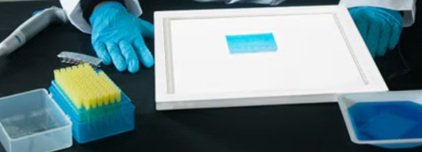
Forensic DNA fingerprinting has become a universally accepted crime-fighting tool. Recent advances use the polymerase chain reaction (PCR) to amplify human DNA obtained from crime scenes. This experiment, based on a crime scene scenario, has an inquiry-based component.
• Learn about DNA fingerprinting while solving a crime!
• Introduce the biotechnologies of restriction enzyme digestion, PCR, and electrophoresis
• Perform agarose gel electrophoresis to separate differently-sized DNA molecules
• Load, run, and analyze Ready-to-Load™ PCR reactions obtained from suspects and the crime scene.
Group Size:
For 8 gels
Time Required:
Complete in 45 minutes
Kit Includes:
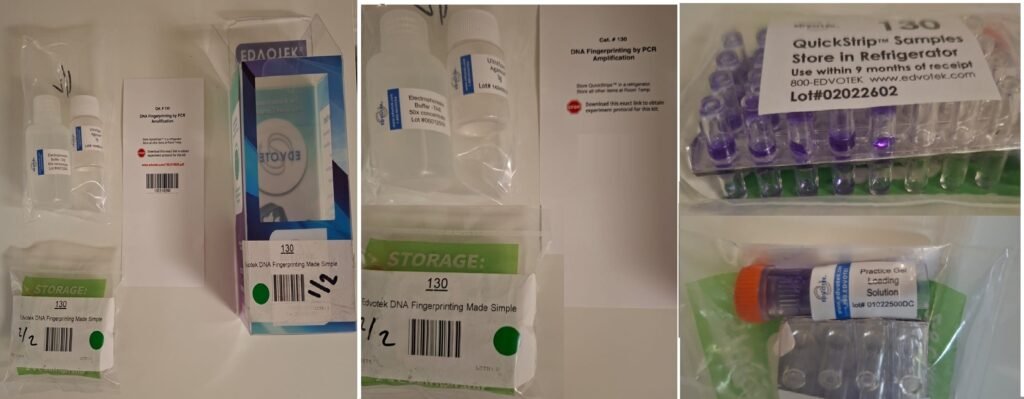
Instructions, Ready-to-Load QuickStrip™ DNA Samples, UltraSpec-Agarose™, Electrophoresis Buffer (50X), Practice Gel Loading Solution, FlashBlue™ DNA Stain.
All You Need:
DNA Electrophoresis, Micropipettes: 5-50 µl (Optional), White Light Box, & Microwave or Hot Plate.
Storage:
Room Temperature Stable. Storage of Ready-to-Load QuickStrip™ samples in the Refrigerator is Recommended.
DNA fingerprinting by PCR amplification is a rapid, highly sensitive technique used in forensics and paternity testing that selectively copies microscopic amounts of trace DNA. By targeting highly variable, non-coding regions—such as Short Tandem Repeats (STRs) or Variable Number Tandem Repeats (VNTRs)—scientists generate billions of exact replicas from a single hair root or saliva trace.
Edvotek Instructional Video: Introducing the TruBlu2 Transilluminator
BioTech Tips: Pipetting – YouTube
1. The Amplification Process (PCR)
- Denaturation (95° C): The template DNA is heated to break hydrogen bonds, separating the double helix into two single strands.
- Annealing (~ 50–65° C): The temperature is lowered to allow short DNA sequences called primers to bind specifically to the targeted STR regions.
- Extension (~ 72° C): A heat-stable enzyme (Taq polymerase) synthesizes new complementary strands, building up the DNA chain.
- Result: Repeating this cycle 30 to 40 times exponentially multiplies the target DNA, producing enough sample for downstream visualization.
2. Visualization & Analysis
- Gel Electrophoresis: The amplified DNA fragments are loaded into an agarose gel matrix and subjected to an electrical current. Because DNA is negatively charged, it migrates toward the positive electrode.
- Size Separation: Shorter STR fragments navigate the gel matrix faster than longer ones, separating the sample into distinct bands.
- Comparison: The banding patterns or fluorescent peaks of the unknown sample (e.g., from a crime scene) are compared against known suspects or reference markers to determine a match.
Key Advantages
- Sensitivity: Requires only trace amounts of starting material.
- Degradation Resilience: Effective even on partially degraded or older samples.
- Speed: Amplification and analysis can typically be completed in a few hours.
Would you like to explore how this method distinguishes between STR analysis versus traditional RFLP (Restriction Fragment Length Polymorphism), or dive deeper into its applications in forensic investigations?