Composition**
Ingredients
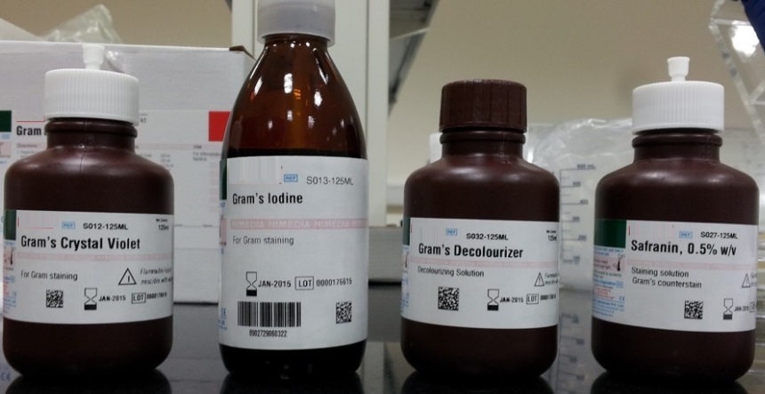
Gram’s Crystal Violet (S012)(Solution A) –
Crystal Violet 2.000 gm
Ethyl alcohol,95% 20.000 ml
Gram’s Crystal Violet (S012)(Solution B) –
Ammonium oxalate 0.800 gm
Distilled Water 80.000 ml
Solution A and B are mixed and stored for 24 hours before use. The resulting stain is stable.
Gram’s Decolourizer(S032) –
Acetone 50.0 ml
Gram’s Iodine(S013) –
Iodine 1.000 gm
Potassium iodide 2.000 gm
Distilled water 300.000 ml
–
0.500 gm
**Formula adjusted, standardized to suit performance parameters
Himedia Gram Stain Kit Directions
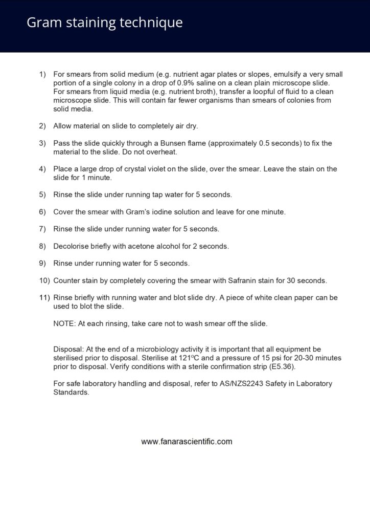
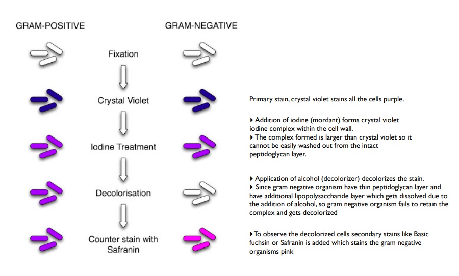
1. Prepare a thin smear on clear, dry glass slide.
2. Allow it to air dry and fix by gentle heat.
3. Flood with Gram’s Crystal Violet (S012) for 1 minute. (If over staining results in improper decolourization of known gram negative organisms, use less crystal violet).
4. Drain the stain.
5. Flood the smear with Gram’s Iodine (S013). Allow it to remain for 1 minute.
6. Decolorize with Gram’s Decolorizer (S032) until the blue dye no longer flows from the smear.
7. Wash with tap water.
8. Counter stain with 0.5% w/v Safranin (S027). Allow it to remain for 1 minute.
9. Wash with water.
10. Allow the slide to air dry or blot dry between sheets of clean bibulous paper and examine under oil immersion objective.
Principle And Interpretation
The Gram stain is a defferential staining technique most widely applied in all microbiology desciplines laboratories. It is one of the most important criteria in any identification scheme for all types of bacterial isolates. Defferent mechanisms have been proposed to explain the gram reaction. There are many physiological defferences between gram-positive and gram-negative cell walls. Ever since Christian Gram has discovered Gram staining, this process has been extensively investigated and redefined. In practice,a thin smear of bacterial cells is stained with crystal violet, then treated with an iodine containing mordant to increase the binding of primary stain. A decolourizing solution of alcohol or acetone is used to remove the crystal violet from cells which bind it weakly and then the counterstain (like safranin) is used to provide a colour contrast in those cells that are decolourized.
Gram-positive bacteria have a thick mesh-like cell wall made of peptidoglycan (50–90% of cell envelope), and as a result are stained purple by crystal violet, whereas gram-negative bacteria have a thinner layer (10% of cell envelope), so do not retain the purple stain and are counter-stained pink by safranin. In a properly stained smear by gram staining procedure, the gram-positive bacteria appear blue to purple and gram negative cells appear pink to red.
Type of Specimen
Any isolated colony on primary or subculture plates can be isolated from following specimens. Clinical specimen: Blood, urine, CSF, pus, wounds, lesions, body tissues, sputum etc. From environment: Air, water, soil, sludge, waste water, food, dairy samples etc.
Specimen Collection and Handling
For clinical samples follow appropriate techniques for handling specimens as per established guidelines.
food and dairy samples, follow appropriate techniques for sample collection and processing as per guidelines. water samples, follow appropriate techniques for sample collection, processing as per guidelines and local standards. Generally the smear is made in laboratory; however, when there is a concern that transport will be delayed or that the preservation for culture will alter the specimen, prepare smear and submit slides to the laboratory.